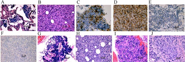
Figure 2. Histology of the primary tumors of patients and patient-derived tumor xenograft (PDTX) tumors. Microphotographs of H&E staining of 010000 in a patient show that the primary tumor (A) and PDTX tumor (B) exhibited the same cell morphologies. Both the primary tumor (C) and PDTX tumor (D) were positive for CD56 staining and negative for TTF1 staining (E and F). Microphotographs of the H&E staining (magnification, ×40) of 010026 in a patient shows that the primary tumor (G) and PDTX tumor (H) exhibited the same cell morphologies. Microphotographs of the H&E staining of 010027 in a patient show that the primary tumor (I) and PDTX tumor (J) exhibited the same cell morphologies.