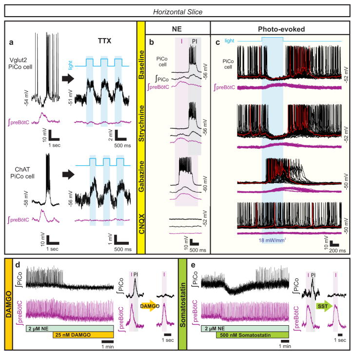
Figure 2. Glutamatergic, cholinergic PiCo cells and role of synaptic inhibition.
a, Intracellular PiCo recordings in Vglut2-cre;Ai27 (n=3) and Chat-cre;Ai27 (n=5) horizontal slices. Photo-stimulation after TTX depolarizes membrane potential. b, PiCo, preBötC in progressive synaptic block (n=5). Rhythms synchronized in gabazine, bursting abolished in CNQX. c, PiCo cell in Dbx1-cre;Ai27 horizontal slice with progressive synaptic block. PiCo cell inhibited during photo-evoked inspiratory burst in NE/strychnine, bursts with light-stimulation in gabazine, ceases bursting in CNQX (500 ms light, 40 sweeps, n=4). d, PiCo bursting eliminated by 25 nM DAMGO; representative bursts (n=5). e, PiCo inhibited by 500 nM SST; representative bursts (n=6).

