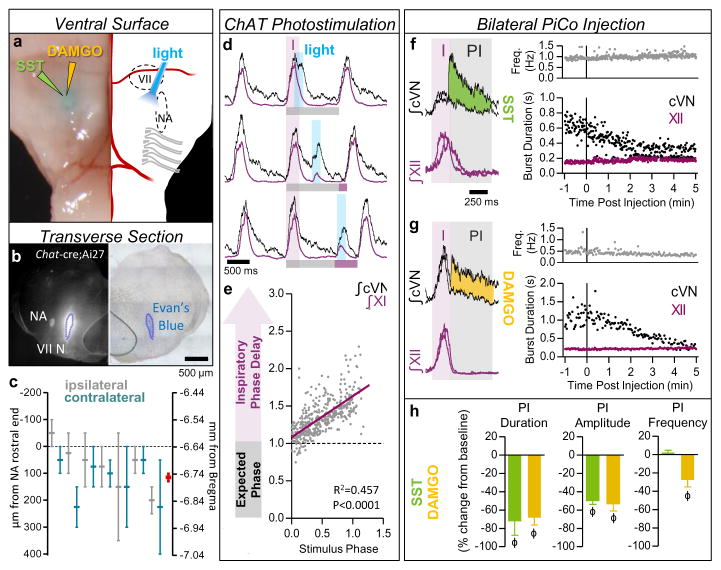
Figure 3. Stimulation and inhibition of PiCo in vivo.
a, Left, brightfield image; right, schematic; ventral sites for bilateral photo-stimulation, injection of SST/DAMGO in vivo. b, Representative dye injection at PiCo level. Left, Chat-cre;Ai27; right, brightfield. c, Dye-spread (n=7), centered at PiCo (0–200 μm caudal from rostral NA). Ipsilateral (grey), contralateral (teal) injections, bars ±max/min; pooled data in red, mean ± s.e.m. d, PiCo photo-stimulation in adult Chat-cre;Ai27 mice evokes vagal bursts and delays subsequent inspiration (grey bars = expected inspiratory phase, purple bars = inspiratory phase delay) e, Quantification of inspiratory phase delay (n=6); magnitude dependent on stimulus phase (slope: 0.549, linear regression analysis). f,g, Injection of SST or DAMGO progressively decreases cVN, not XII burst duration. h, Postinspiratory burst duration, amplitude, and frequency following injection of SST (n=3), DAMGO (n=4). Two-tailed paired t-test, ϕ P<0.05 compared to baseline; mean ± s.e.m.

