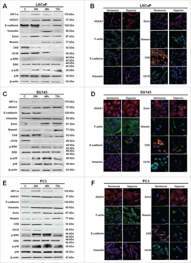
Figure 2.

Western blot using antibodies against HIF1-α, ANXA1, E-cadherin, vimentin, ezrin, moesin, CK8, CK18, p-ERK, ERK, p-p38, p38 on protein extracts from LNCaP (A), DU145 (C) and PC3 (E) cells. Protein normalization was performed on β-actin levels. Immunofluorescence analysis to detect: ANXA1, F-actin, E-cadherin, vimentin, ezrin, moesin, CK8 and CK18 on LNCaP (B), DU145 (D) and PC3 (F) cells in both normoxia and hypoxia conditions. Immunofluorescence images refer to 48 hours of low oxygen treatment. Nuclei were stained with DAPI. Magnification 63x. Bar = 10 μm. The data are representative of 3 experiments with similar results.
