
Figure 2. CobB deacetylates MDHs.
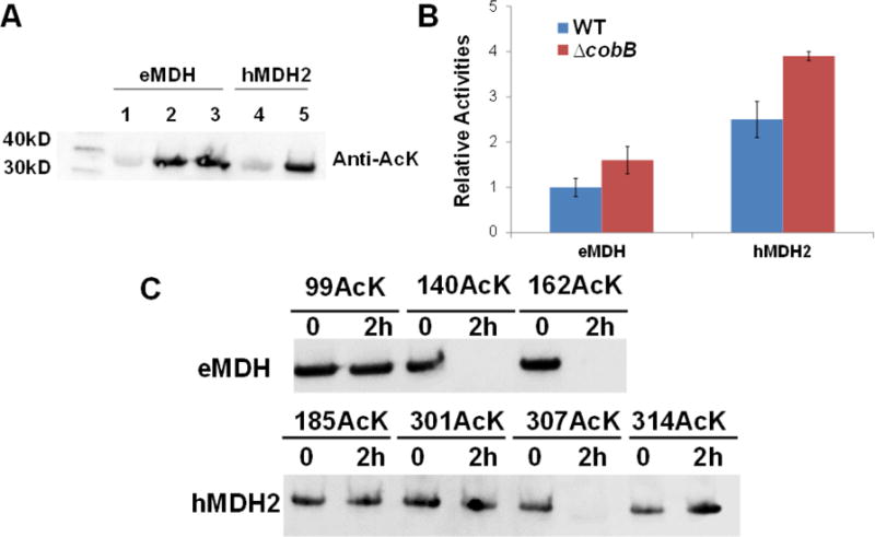
A) Western blotting of purified MDHs from TOP10 and TOP10 ΔcobB cells. Lane 1 and 4 were from wild-type TOP10 cells. Lane 2 and 5 were from TOP10 ΔcobB cells. Lane 3 was from TOP10 ΔcobB ΔyfiQ cells. The same amounts of proteins were loaded. B) The enzyme activities of purified MDHs from TOP10 and TOP10 ΔcobB cells. The activity of eMDH purified from wild-type TOP10 cells was set as 1. The mean values and standard errors were calculated based on three replicates. C) Western blotting of acetylated MDH variants treated with the CobB protein. The acetylation levels of MDH variants after 2-hour incubation were compared with those without CobB treatment. The same amounts of proteins were loaded.