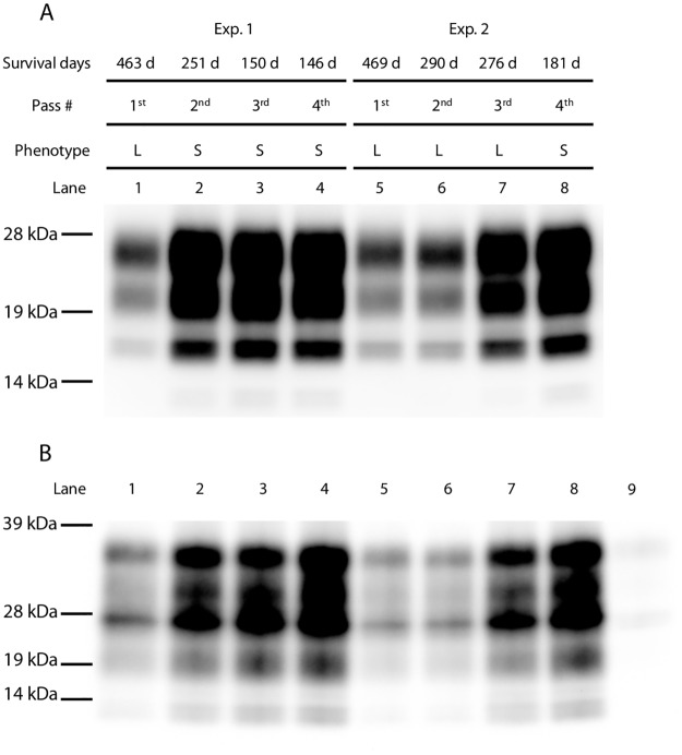
Fig 2. Monitoring PrPSc accumulation in diseased mice throughout passaging.
(A) Representative western blot of PrPSc in mouse brains at different passages. Experiment number, survival days, passage number, disease phenotype, and lane number are indicated on the top of the panel. Each lane included 0.2 mg brain equivalents. (B) Immunoprecipitation assay with mAb 3H6. Lane number is indicated on the top of the panel. In lane 9, uninfected brain homogenate was probed with mAb 3H6. In all other lanes, PrPSc was detected with mAb T2-HRP. Molecular markers are shown on the left side of each panel.

