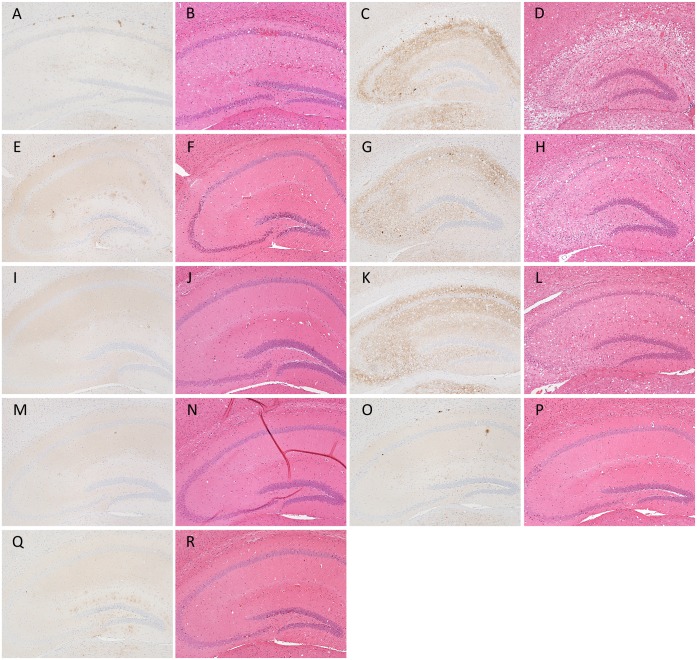
Fig 3. Representative images of PrPSc deposition types associated with each prion phenotype in ICR mice.
Sections of the hippocampus were subjected to immunostaining of PrPSc and H&E staining. Typical PrPSc distribution patterns and histopathology of the hippocampus affected with L-type (A and B) and S-type (C and D) prions are shown. Sections of the hippocampi of mice inoculated with cell homogenates prepared from GT/Mo1 (E and F), GT/Mo2 (G, H, I and J), GT/Mo3 (K and L), GT/Mo1 ′ (M and N), GT/Mo2′ (O and P), and GT/Mo3′ (Q and R) were also subjected to immunostaining of PrPSc and H&E staining. Immunohistochemical detection of PrPSc was performed using the mAb SAF84.

