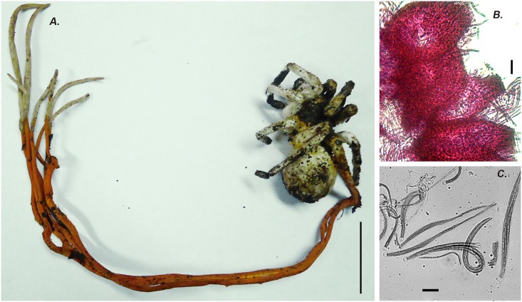
Fig 3. Cordyceps caloceroides.
A) A long stromata emerging from a big Theraphosidae spider (picture taken by Carlos Padilla on Yasuni Scientific Research Station, EC). B) Cross section of perithecia stained with Fucsin lactic acid showing the broad ostioles HUA 186166. C) Asci stained with cotton blue dye showing the curved filiform ascospores Scale bars: A = 5 cm, B = 10 μm 40 ×, C = 10 μm 100 ×.

