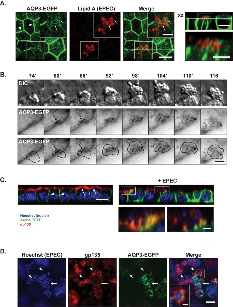
Fig 2. Basolateral AQP3-EGFP localized to the center of EPEC microcolonies, whereas apical gp135 localized to the periphery.
A. MDCK-AQP3-EGFP cells were seeded to confluency on semi-permeable collagen-coated Transwell filter supports and allowed to polarize for 3 days. Cells were then infected with EPEC for 6 hours, fixed and stained with an antibody against Lipid A to label EPEC bacteria (shown in red). The rightmost image shows a xz view at the position indicated by the white line in the merge image. AQP3-EGFP localized to the lateral membrane (arrow heads) and also accumulated at the site of microcolony formation around individual EPEC bacteria (arrows). Scale bars: 10 μm and 5 μm (insets). B. MDCK-AQP3-EGFP cells were infected with EPEC bacteria directly into the heating chamber after mouting on the microscope. Time-lapse imaging was performed with 1 minute intervals for DIC (EPEC and cells) and EGFP (AQP3-EGFP). Montage shows DIC and AQP3-EGFP in inverted contrast. The bottom panel shows the EGFP image including a drawn outline of the bacterial microcolony based on the DIC image. AQP3-EGFP recruitment was observed at the center of microcolony formation (at approximately 80’ after initial attachment) and was sustained to the center of the microcolony with no detectable recruitment to the periphery of the EPEC colony. Scale bar: 5 μm. C-D. Polarized MDCK-AQP3-EGFP monolayers with and without EPEC infection for 6 hours were stained with a monoclonal anti-gp135 antibody (red) and hoechst (blue, to detect EPEC and cell nuclei). C. xz-projection of confocal z-stacks showed that AQP3-EGFP was localized to the basolateral membrane (white arrow heads), whereas gp135 was strictly localized to the apical membrane (white arrows) in the uninfected cells. Both AQP3-EGFP and gp135 were localized at the infection site in EPEC-infected cells. Scale bar: 10 μm and 3 μm for insets. D. xy maximum projection of 6 slices from a z-stack showing an EPEC microcolony. Arrows point to AQP3-EGFP, arrowheads to gp135. Gp135 seemed less intense at sites of AQP3-EGFP accumulation. Scale bar: 10 μm and 1 μm for the inset.

