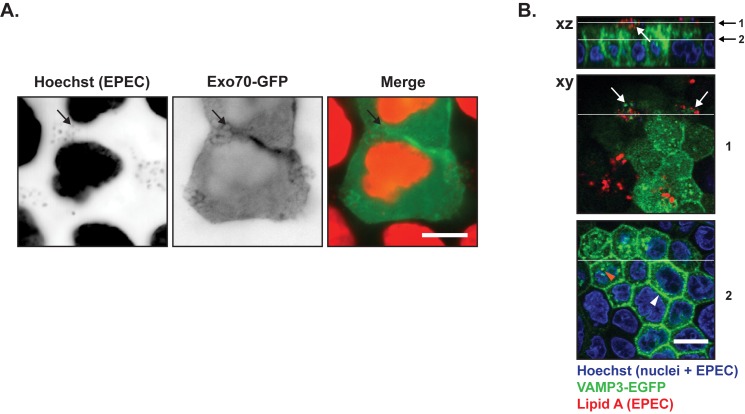
Fig 4. Components of the basolateral vesicle docking/fusion machinery were recruited to EPEC microcolonies.
A. Subconfluent MDCK cells were transiently transfected with Exo70-GFP, which is part of the basolateral exocyst vesicle docking hub. The cells were infected with EPEC for 4 hours, fixed and stained with hoechst to label cell nuclei and EPEC bacteria. Hoechst labeling and Exo70-GFP are shown as inverted contrast and in red and green in merge, respectively. Arrows indicate examples of EPEC bacteria with Exo70-GFP localization. Scale bar: 10 μm B. Polarized MDCK-VAMP3-EGFP cells on semi-permeable Transwell filters were infected with EPEC for 6 hours, fixed and stained with anti-Lipid A to label EPEC bacteria (red) and hoechst to label cell nuclei as well as EPEC bacteria. In the basal region of the cell, VAMP3-EGFP localized at cell-cell contacts (white arrowhead) and as puncta in the cytosol (orange arrowhead). In the apical part of the cells,VAMP3-EGFP was concentrated at the sites of infection (white arrows). Positions of xy sections and xz projections are indicated by white lines, 1 is through an EPEC colony, 2 is close to the plasma membrane. Scale bar: 10 μm.

