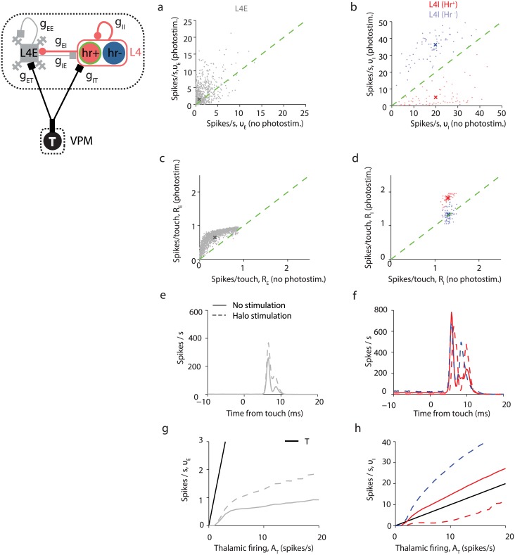
Fig 11. Simulated light activation of halorhodopsin expressed in L4I-Hr+ neurons.
Simulations with fhalo = 0.5 reveals a reduction in the whisking suppression and an enhancement of touch responses by L4E neurons. (a) Halorhodopsin activation in L4I-Hr+ causes an average increase in response of L4E during whisking and no touch, with a wide distribution of halorhodopsin—induced modifications. (b) Most L4I-Hr+ neurons reduce their activity during whisking while L4I-Hr- neurons increase it. (c) Increase in the touch responses in L4E neurons during suppression of L4I-Hr+. (d) Increase in the touch responses in L4I neurons. The increase in touch responses is only seen in Hr+ cells. (e,f) Population PSTH of L4E (e) and L4I (f) neurons with and without L4I-Hr+ activity suppression. (g) Reduction of L4I-Hr+ activity diminishes the whisking suppression effect in L4E neurons. Black line: T neurons; solid grey line: L4E neurons without halorhodopsin activation; dashed grey line: L4E neurons during halorhodopsin activation. (h) Reduction of L4I-Hr+ activity diminishes the whisking response in L4I-Hr+ neurons. Solid red line: L4I-Hr+ neurons without halorhodopsin activation; dashed red line: L4I-Hr+ neurons during halorhodopsin activation. Dashed blue line: L4I-Hr- neurons during halorhodopsin activation.

