
Figure 1.
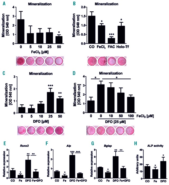
Iron excess inhibits and iron chelation supports osteogenic differentiation. (A–D) Bone marrow stromal cells were differentiated towards osteoblasts in the presence of (A) various concentrations of iron chloride (FeCl3), (B) 25 μM FeCl3, 1 μM ferric ammonium citrate (FAC), and 250 μM holo-transferrin (Holo-Tf), (C) the iron-chelator deferoxamine (DFO) as well as (D) combinations of FeCl3 and DFO for 21 days. The mineralization was visualized with alizarin red S staining and quantified after elution with cetylpyridinium chloride. (E–G) Gene expression analysis of Runx2, alkaline phosphatase (Alp) and osteocalcin (Bglap) using real-time polymerase chain reaction (PCR) after treating day 10 differentiated cells with 5 μM FeCl3 (Fe), 25 μM DFO or Fe+DFO for an additional 48 hours. (H) Enzyme activity of ALP in day 10 osteoblasts after 48 hours of Fe (5 μM) and DFO (25 μM) treatment. N=4–6. *P<0.05, **P<0.01, ***P<0.001 vs. control (CO).