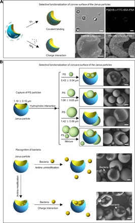
Fig. 4. Selective surface functionalization of the Janus particles.

(A) Selective functionalization of the convex surface of the Janus particles. The fluorescent FITC-BSA molecules were selectively modified onto the convex surface of the PSDVB ﬤ PAA Janus particles. Scale bar, 2 μm. Meanwhile, inorganic nanoparticles, such as Ag and Fe3O4 nanoparticles, were also locked onto the convex surface of the Janus particles via one-step electrostatic attraction. Scale bar, 500 nm. (B) Selective functionalization of the concave surface of the Janus particles. Janus particles were used to capture and recognize spherical PS particles and live bacteria (S. aureus). When the PSDVB ﬤ PAA Janus particles were mixed with spherical PS particles with diameters of 0.43 ± 0.04 μm, 1.08 ± 0.05 μm, and 1.42 ± 0.06 μm, respectively, those with diameters of 0.43 ± 0.04 μm and 1.08 ± 0.05 μm were captured on the concave surfaces, whereas those with diameters of 1.42 ± 0.06 μm were difficult to capture. When the PSDVB ﬤ PAA Janus particles were simultaneously mixed with PS particles with three different particle sizes, the PS spheres with average diameters of 1.08 ± 0.05 μm were the more easily captured particles. Meanwhile, the concave surfaces of the Janus particles functionalized by aniline were used to selectively recognize live bacteria through electrostatic interactions. Scale bar, 500 nm.
