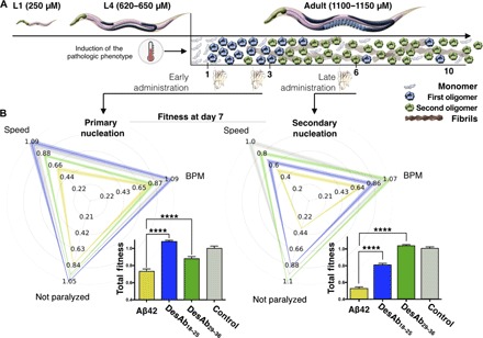
Fig. 4. Effects of DesAb18–25 and DesAb29–36 in a C. elegans model of Aβ42-mediated toxicity.

(A) Experimental design for the investigation of the effects of the two selected DesAbs in the C. elegans strain GMC101 (the Aβ42 worm model) compared with strain N2 (the control worm model). The pathological phenotype is induced in the worms by increasing their temperature of incubation from 20° to 24°C, which induces Aβ42 aggregation. A pictorial representation of the populations of monomers (light blue), oligomers formed by primary (blue) and secondary (green) nucleations, and fibrils (maroon) at the different stages (in days) of adulthood of the worms is given to illustrate the aggregation process. (B) Phenotypic fingerprints, which consider speed, body bends per minute (BPM), and fraction not paralyzed or the worms, of Aβ42 worms (C. elegans GMC101; yellow) and control worms (C. elegans N2, WT; gray) treated with empty lipid vesicles and after the administration of DesAb29–36 (green) and DesAb18–25 (blue), screened at day 7 of adulthood. DesAbs were administered starting from a 20 μM concentration (see Materials and Methods) at days 1 and 3 (left) or at day 6 (right). The fingerprints show one representative of three biological replicates that showed similar results. The thickness of the lines represents SEM. The bar plots report the total fitness (see Materials and Methods). *P ≤ 0.05, **P ≤ 0.01, ***P ≤ 0.001, and ****P ≤ 0.0001 (relative to untreated worms).
