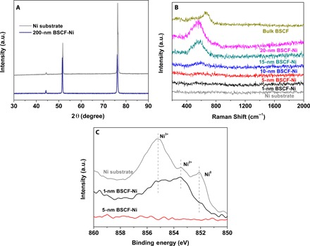
Fig. 1. Characterization of amorphous BSCF nanofilms on Ni foil substrates.

(A) Powder XRD patterns for the 200-nm BSCF–Ni foil and the Ni foil substrate. (B) Raman spectra for the 1-, 5-, 10-, 15-, and 20-nm BSCF on Ni foil as well as the Ni foil substrate and BSCF particle for reference. (C) Ni 2p XPS for the Ni foil as well as 1- and 5-nm BSCF–Ni foil heterostructures.
