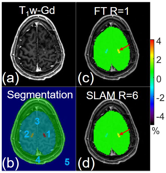
Figure 4.

(a) Post-gadolinium T1-weighted (T1w-Gd) image and (b) 5-compartment segmentation overlaid on the T1w-Gd image. Color-coded APT-weighted images (c) from the standard compartmentally-averaged FT method and (d) from SLAM with an acceleration factor of 6. Both FT and SLAM were processed in a slice-by-slice fashion.
