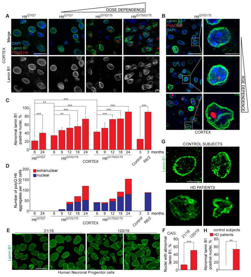
Figure 1. Expanded polyQ Huntingtin disrupts neuronal nuclear envelope morphology in a dose- and age-dependent manner.
(A) Immunofluorescence (IF) of lamin B1 (green) and polyQ Htt (red) in cortex of mice at indicated ages expressing one (HttQ7/Q175) or two (HttQ175/Q175) copies of expanded polyQ Htt and wild type littermates (HttQ7/Q7). (B) IF of lamin B1 (green) and polyQ Htt (red) in cortex of aging HttQ7/Q175 mice. A, B: Nuclei were stained with DAPI. (C) Percentage of altered nuclear envelopes in cortical sections of mice of indicated genotypes and ages, as determined by IF of lamin B1. 100–400 nuclei were counted per genotype and age. (D) Quantification of nuclear and extranuclear aggregates of polyQ Htt in cortex of HttQ175 and R6/2 mice. 200 cells were counted per genotype and age. (E, F) IF of lamin B1 (E) and percentage of altered nuclear envelopes (F) in human iPSC-derived neuronal progenitor cells with indicated number of CAG repeats. At least 600 cells were analyzed for each genotype. (G, H) IF of lamin B1 (G) and percentage of altered nuclear envelopes (H) in sections of motor cortex of two non-neurological disease control subjects and two Huntington’s disease patients of indicated ages. At least 200 nuclei were counted per each group. A, B, G: Z projections (3 μM) are shown. C, F, H: Data are shown as mean ± SEM. **: P<0.01, ***: P<0.001, chi-square test.
See also Figures S1 and S2.

