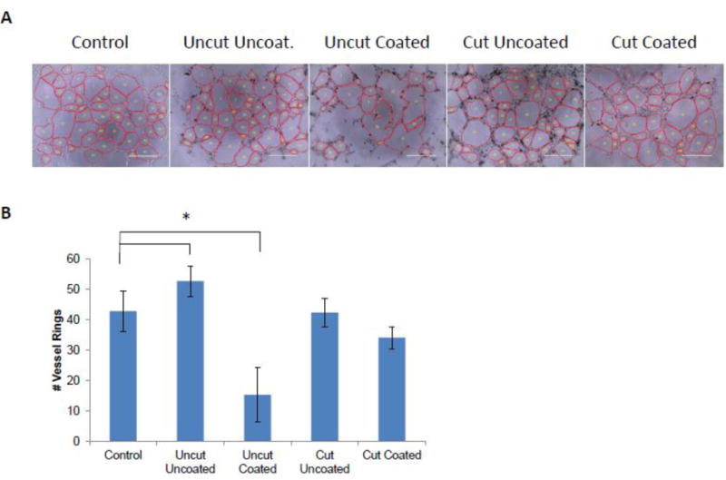
Figure 3. Tube Formation Assay of MWCNT-treated HBMECs.

HBMECs were treated in monolayer overnight with 25 μg/mL of each type of MWCNTs and then were replated on matrigel to induce tube formation. After 6 h, photomicrographs of tube formation were taken. Representative images are shown in (A). The average number of HBMEC vessel rings was quantified in three independent biological replicates, with each biological replicate containing triplicate technical replicates. Data are displayed as the mean ± standard error of each experiment (B). Significant differences among groups are indicated: *p<0.05 (ANOVA; post-hoc T-Test).
