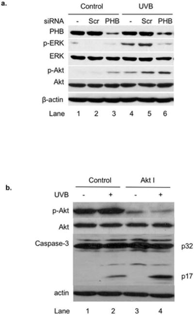
Figure 4.

The role of PHB in UVB-induced apoptosis in HaCaT cells. (a) HaCaT cells were transiently transfected with scrambled siRNA or PHB siRNA. The cells without transfection were used as control. The levels of PHB, p-ERK, ERK, p-Akt, Akt, and β-actin in the transfected cells with or without UVB (50 mJ/cm2, 6 h) were assessed by western blotting analysis. The data represents three experiments. (b) The levels of p-Akt, Akt, caspase-3, and β-actin in Akt I (1 μM) and/ or UVB (50 mJ/cm2, 6 h) treated cells were assessed by western blotting analysis. The data represents three experiments.
