
Fig. 1.
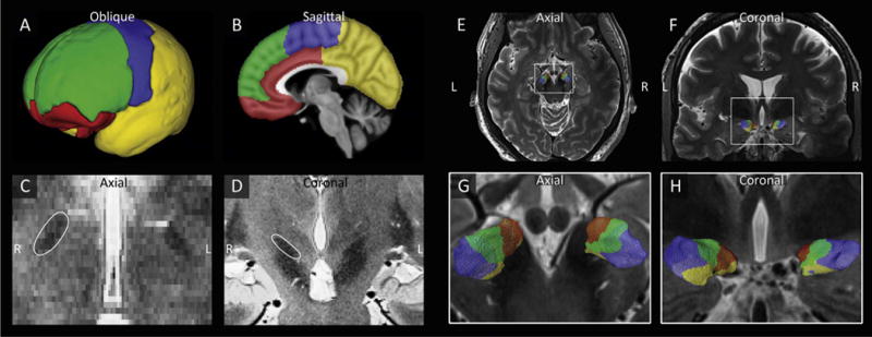
The STN is parcellated based on its connections to the limbic, associative, motor, and remaining cortical areas. A–B) Division of the cortex into limbic (red), associative (green), motor (blue) and remaining (yellow) cortical areas. C–D) Visualization of the hypointense STNs in the axial (C) and coronal (D) planes. E–H) Example of the parcellation of the STNs of one subject in axial (E,G) and coronal (F,H) views.