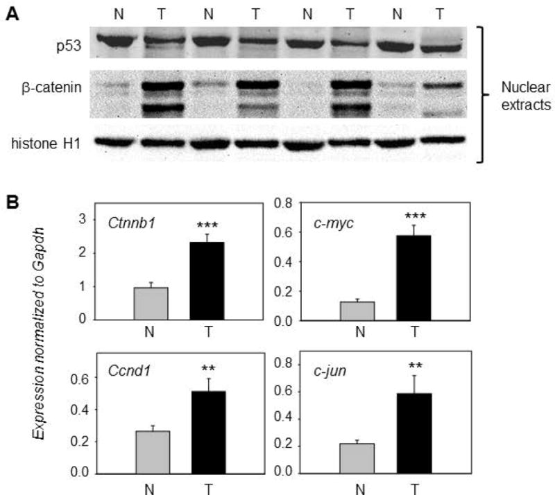
Figure 2.

Overexpression of β-catenin in PhIP-induced skin tumors. (A) Representative immunoblots of β-catenin and p53 in nuclear fractions; histone H1, loading control; T, tumor; N, normal-looking tissue adjacent to tumor. An additional set of five T/N pairs generated similar data for the proteins indicated (immunoblot data not presented). (B) qRT-PCR analyses of selected β-catenin target genes normalized to glyceraldehyde-3-phosphate dehydrogenase (Gapdh); mean±SD, n=12, **P<0.01, ***P<0.001. Each panel is representative of an experiment that was repeated at least three times, for each gene mentioned, using twelve matched pairs of T/N samples.
