Figure 3.

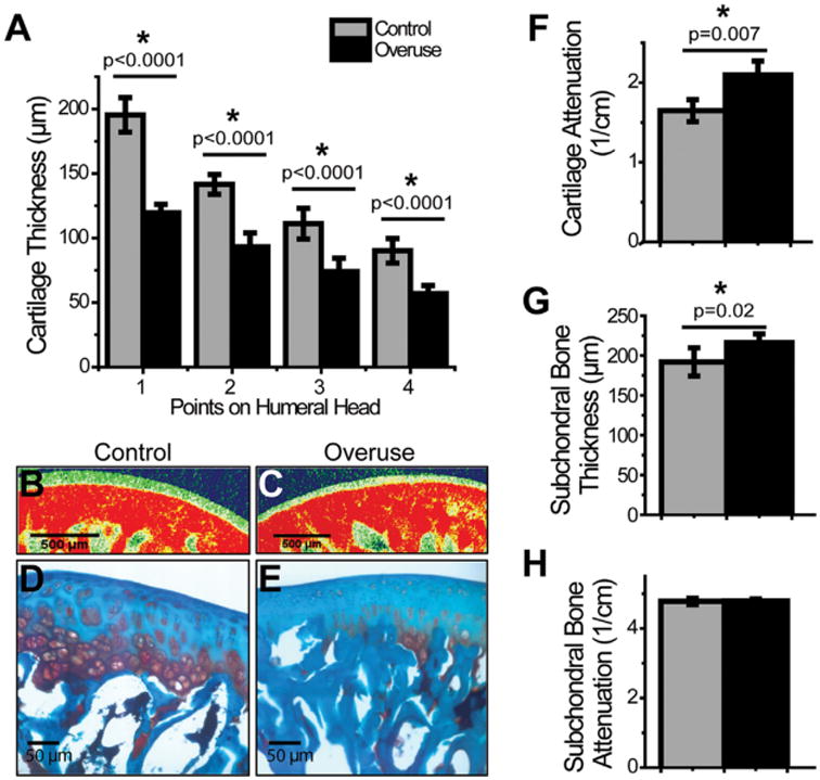
EPIC-μCT measurements of humeral articular cartilage and subchondral bone. Humeral heads demonstrated a reduced cartilage thickness in overuse animals in comparison to their age-matched controls (A). Humeral cartilage also displayed decreased GAG content by EPIC-μCT in overuse animals in comparison to controls (B, C, F) (SD: control: ± 0.14cm−1; overuse: ± 0.18 cm−1), which was confirmed with safranin-O staining of the decalcified humeral head (D and E). While subchondral bone thickness was higher in overuse animals as compared to controls (G), there was no difference in mineral attenuation in the subchondral bone (H). *Denotes significant difference, p values as stated (n = 8 ± SD).
