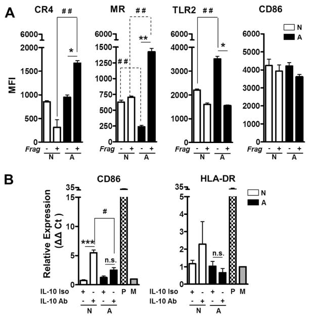
Fig. 3. Effects of ALF fragments on macrophage receptors and activation.
(A) Macrophages exposed to ALF exposed M.tb [A] or 0.9% NaCl exposed M.tb [N] released cell wall fragments induce the surface expression of the MR and CR4, but not TLR2. Differences in the macrophage activation marker CD86 on the macrophage surface were not observed. Experiments were performed n=3 and cells analyzed by flow cytometry. Shown M±SEM, Student’s t test was used to compare the receptor/marker surface expression induced by fragments generated by M.tb exposure to ALF with fragments generated by M.tb exposure to NaCl (*P<0.05; **P<0.005). (B) Expression of CD86 and HLA-DR as measured by RT-PCR (n=3). P: Positive Control; M: Medium. Student’s t test was used to compare the mRNA levels induced by fragments generated by M.tb exposure to ALF with fragments generated by M.tb exposure to NaCl in the presence (#P<0.05; ***P<0.0005) or in the absence of anti-IL-10 Ab. For each ‘n’ value, both ALF and macrophages were obtained from different human donors.

