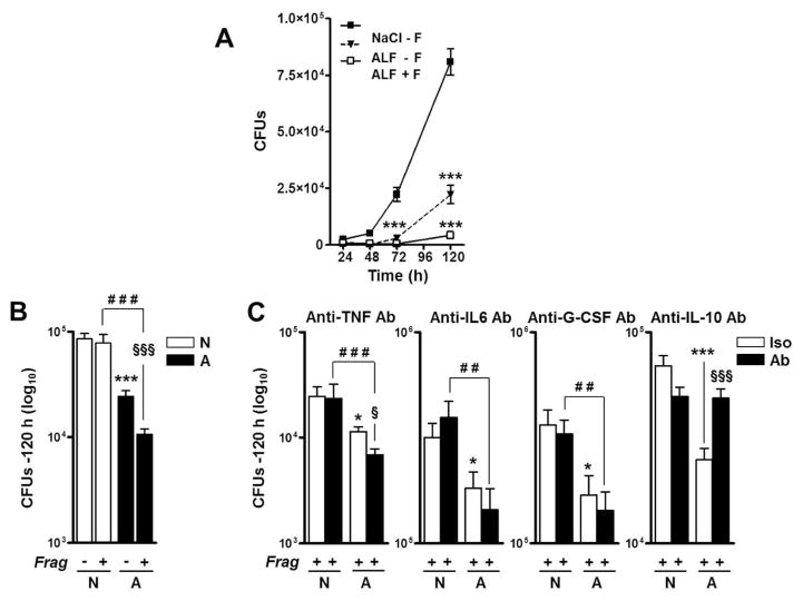
Fig. 4. Effects of ALF on M.tb intracellular survival in human macrophages.
M.tb bacilli were exposed to 0.9% NaCl (control), Mix, or ALF at relevant in vivo concentrations. Macrophages were infected at a MOI 1:1 with exposed M.tb in the absence or presence of fragments and in the absence or presence of anti-TNF, anti-IL-10, anti-IL-6 or anti-G-CSF neutralizing Abs. Intact macrophage monolayers were verified by inverted phase microscopy throughout the infection period. M.tb survival was determined by CFUs at the indicated intervals. (A) A representative experiment in triplicate is shown (M±SD), Student t test was used to compare control with vs. without fragments (±F), ***p<0.0005. (B) Overall data at 120 h from n=3 each performed in triplicate (M±SEM). (C) Effects of neutralizing TNF, IL-10, IL-6 or G-CSF on macrophages in the presence of fragments during exposed M.tb infection (n=3 in triplicate, M±SEM). Notice that y-axes are different among the graphs shown. One-way ANOVA, Tukey-Posttest was used to compare the effects on the M.tb cell wall (*, white bars), ± F (§, white vs. black bars), and M.tb exposed to different conditions in the presence of fragments (#, black bars), *P<0.05; **P<0.005; ***P<0.0005; §P<0.05; §§§P<0.0005; ##P<0.005; ###P<0.0005. NaCl-M.tb + Frag [N]; ALF-M.tb + Frag Frag [A]. For each ‘n’ value, both ALF and macrophages were obtained from different human donors.

