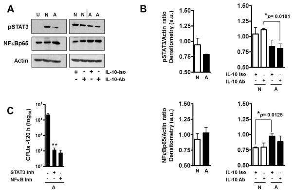
Fig. 6. Effects of ALF on macrophage STAT3 and NFκB activation.
Following infections as described in Fig.4, lysates from ALF-M.tb [A] or NaCl-M.tb [N] infected macrophages in the presence of their respective fragments were obtained. (A) Representative Western blots show that ALF-exposed M.tb in the presence of fragments decreased the activation of STAT3 signaling pathway and increased NFκB activation. This was not an IL-10 dependent mechanism. U: Uninfected; N and A: NaCl- or –ALF-exposed M.tb in the presence of fragments, respectively. (B) Densitometry analysis of n=3 showing phosphorylated STAT3 and NFκB vs. Actin ratios. Student t test was used to compare ALF exposed M.tb with NaCl-exposed M.tb (control) in the presence of fragments, §§P<0.005 (between white and black bars under the same condition). (C) Inhibition of STAT3 and NFκB both allowed macrophages a further enhanced control of ALF-M.tb infection in the presence of fragments. Student t test was used to compare ALF exposed M.tb infection in the presence of fragments in the absence or presence of inhibitors, **P<0.005. For each ‘n’ value, both ALF and macrophages were obtained from different human donors.

