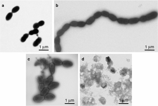
Fig. 5.

TEM analysis with negative staining of S. pneumoniae R6 cells cultured in the presence or absence of the Natto peptide. The cells were cultured in TH broth containing 5% (v/v) horse hemolysate in the absence (a, c) or presence (b, d) of the Natto peptide (64 μg/mL). After 4 h (a, b) or 8 h (c, d) of incubation, the cells were harvested and observed TEM after negative staining
