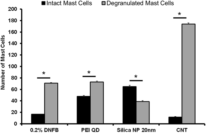
Figure 8.

Mast cells quantified in the ear tissue by IHC using the Geimsa stain. The number of degranulated MCs was significantly lower in the silica NP (20 nm) treatment group compared to PEI-QD and CNT treated ears co-challenged ear. MCs were counted in 10 fields of view at 40X for 3 independent mouse skin samples. Mean ± SEM, *p < 0.05, 2-tailed Student’s t-Test, paired, with respect to intact mast cells in each group. Error Bars: Standard Dev.
