Figure 3.

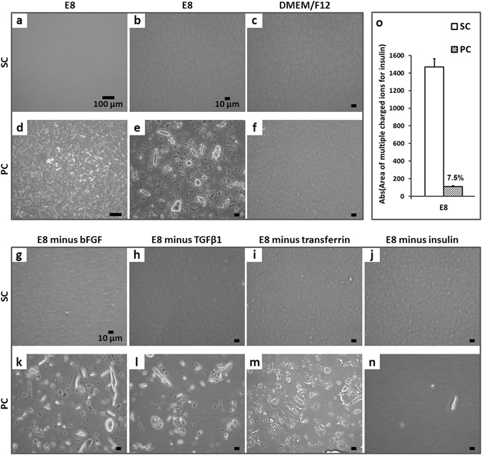
Optical inspection of conditioned media. Light microscopy images of SC E8 at (a) 10x (scale bar = 100 µm) and (b) 40x magnification (scale bar = 10 µm) revealed a transparent medium, whereas in (d–e) PC E8 the presence of irregular particles of up to ~50 µm was observed. The (c) static and (f) peristaltic pump conditioning of DMEM/F12 did not indicate any release of particles from circuit components (scale bar = 10 µm). Similarly, (g–j) no particles were detectable in any SC E8 media lacking one protein at a time omitted before conditioning (scale bar = 10 µm). In contrast, peristaltic pump conditioning resulted in substantial particle formation in (k) E8 minus bFGF, (l) E8 minus TGFβ1, and (m) E8 minus transferrin, except in (n) E8 minus insulin (scale bar = 10 µm). (o) Semi-quantitative UPLC-MS analysis of SC E8 and PC E8 media revealed that less than 10% of dissolved insulin remained in PC E8 compared to SC E8 controls (n = 2).
