
Figure 1.
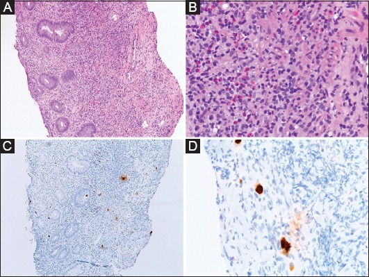
(A) Low magnification (10×), hematoxylin & eosin stain, showing fragments of severely ulcerated colon mucosa. The tissue consists of a mixture of fibrinopurulent exudate and an inflammatory infiltrate that includes lymphocytes, neutrophils, eosinophils and plasma cells. (B) High magnification (40×) multiple cells, showing enlarged nuclei with an intra-nuclear magenta inclusions. (C, D) Immunohistochemical staining (10×, 40×) for cytomegalovirus is positive on the intranuclear inclusions