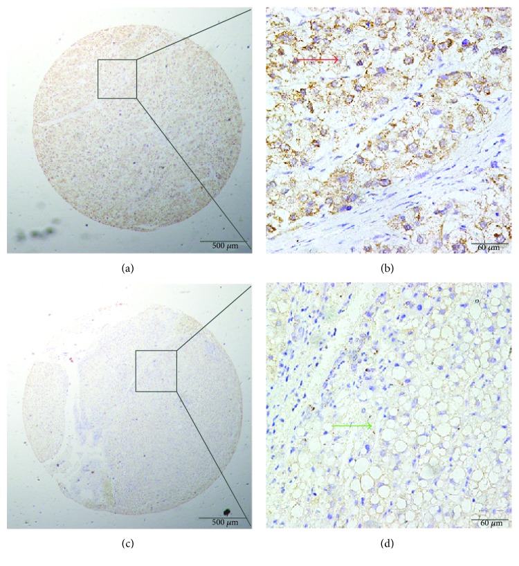
Figure 2.
Immunohistochemical (IHC) staining for Eg5 expression in cancerous and peritumoral tissues. (a, b) Positive cytoplasmic IHC staining (red arrow) for Eg5 in HCC tissue samples. (c, d) Negative IHC staining (green arrow) for Eg5 in normal tissue samples (original magnification: ×40 in (a) and (c); ×400 in (b) and (d)).

