
Figure 1.
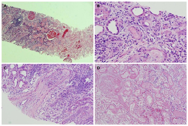
Renal pathology. Pathological findings: A: Corticomedullary junction. The right side is necrotic renal cortex. On the left side of the image there is still viable corticomedullary junction. H and E, × 40; B: An arteriole obliterated with amorphous material (platelet and fibrin thrombus). H and E, × 200; C: An arcuate artery with almost completely obliterated lumen by severe fibrous to mucoid intimal thickening at the corticomedullary junction. H and E, × 100; D: Necrotic renal cortex without nuclear staining in the cells. H and E, × 200.