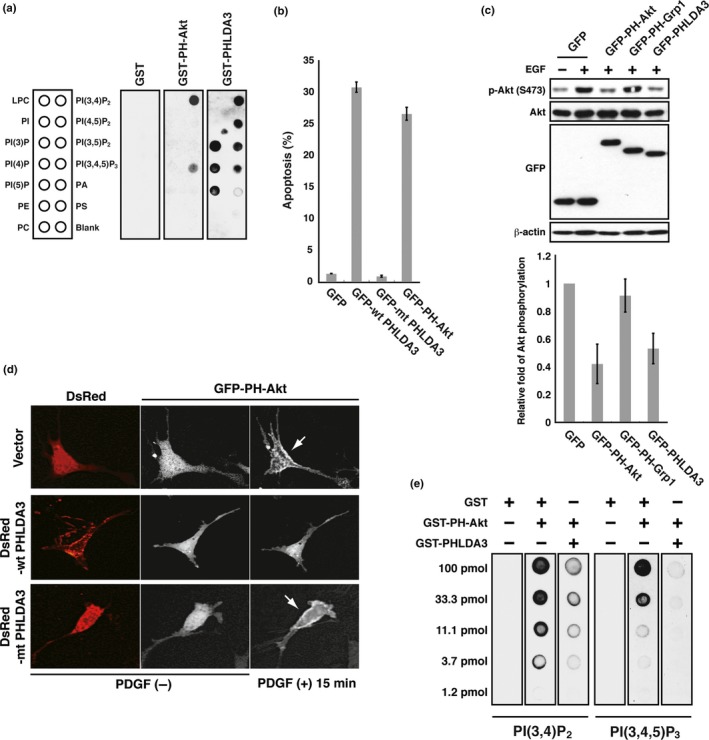
PHLDA3 competes with the PH domain of Akt. (a) Binding of GST‐PHLDA3, GST‐PH‐Akt or GST to immobilized PIP was assessed by protein‐lipid overlay assay. Nitrocellulose membranes spotted with 100 pmol of different phospholipids were used. Bound proteins were detected with anti‐GST antibody. Note that GST alone produced no signal under the conditions employed. LPC, lysophosphatidylcholine; PA, phosphadic acid; PC, phosphatidylcholine; PE, phosphatidylethanolamine; PS, phosphatidylserine. (b) 293T cells were transfected with GFP, GFP‐WT PHLDA3, GFP‐mtPHLDA3 (a PHLDA3 mutant with a small deletion within the PH domain), or GFP‐PH‐Akt and analyzed for GFP‐positive cells 48 h post‐transfection. The apoptotic rate, measured by PI‐positive cells (cells stained with PI without fixation), is shown. Mean apoptotic rates ± SD from three experiments are shown. (c) PHLDA3 inhibits Akt activation. COS7 cells were transfected with the indicated fusion proteins for 24 h and subsequently stimulated with EGF for 5 min. Induction of Akt phosphorylation upon EGF treatment was detected in control cells expressing GFP. Akt activity after EGF treatment was analyzed by western blotting, and Akt activity relative to the GFP‐transfected control was calculated. The mean ± SD from three experiments is shown. GFP fusion protein levels were also analyzed by western blotting. (d) Akt translocation to the plasma membrane upon PDGF treatment was analyzed by live‐cell imaging. NIH 3T3 cells were transfected with GFP‐PH‐Akt together with DsRed, DsRed‐WT PHLDA3 or DsRed‐mtPHLDA3. GFP‐PH‐Akt subcellular localization was monitored before and after PDGF treatment (15 min). Note that Akt is localized at the plasma membrane in cells expressing DsRed or DsRed‐mtPHLDA3 (shown by arrows). (e) PHLDA3 inhibits PH‐Akt binding to PIP
2 and PIP
3. Binding of GST‐PH‐Akt to immobilized PIP was assessed by protein‐lipid overlay assay. Nitrocellulose membranes spotted with serially diluted PIP
2 and PIP
3 were incubated with the indicated proteins. While GST did not interfere with Akt binding to PIP, PHLDA3 significantly interfered. Bound Akt was detected with anti‐Akt PH domain antibody.

