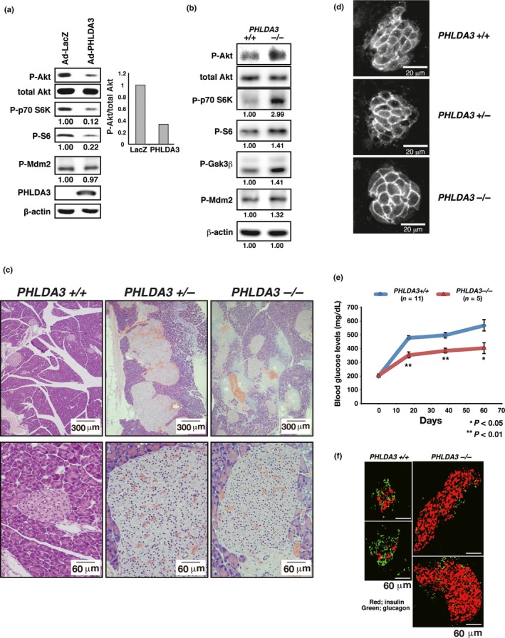
Figure 4.

PHLDA3 function in islet cells. (a) Effect of PHLDA3 expression on Akt activity in MIN6 cells. MIN6 cells were transduced with Ad‐LacZ or Ad‐PHLDA3 at a moi of 35, and harvested 30 h post‐infection. Akt activation and phosphorylation of Akt downstream signaling molecules were analyzed by western blotting and quantified by normalization to total Akt levels (P‐Akt) or by β‐actin levels (P‐p70 S6K, P‐S6, P‐Mdm2). (b) Akt activation and phosphorylation of Akt downstream signaling molecules were analyzed by western blotting and quantified by normalization to total Akt levels (P‐Akt, Right) or by β‐actin levels (P‐p70 S6K, P‐S6, P‐GSK3β and P‐Mdm2). (c) HE staining of islets from wild type, heterozygote and PHLDA3‐deficient 10‐month‐old mice. (d) Islet cell size in wild type, heterozygote and PHLDA3‐deficient mice. (e) Blood glucose levels in streptozotocin‐induced diabetic mice. Indicated numbers (n) of PHLDA3 +/+ or PHLDA3 −/− mice were injected i.p. with streptozotocin (STZ) for 5 consecutive days. Blood glucose levels were determined at different time points as indicated after administration of STZ. (f) Distribution of β and α cells in STZ‐treated PHLDA3 +/+ and PHLDA3 −/− mice. Sections were stained with antibody against insulin (β cell marker; red) and glucagon (α cell marker; green) and representative images are shown.
