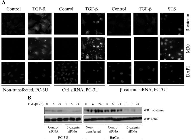
FIG. 6.
Transfection of siRNA for β-catenin reduces TGF-β induced apoptosis. (A) PC-3U and HaCaT cells were transfected or not with specific or unspecific (ctrl) siRNA for β-catenin and treated with TGF-β for 24 h. The localization and amount of β-catenin and number of apoptotic cells, shown by M30 staining, were examined by immunofluorescence. Nuclei were counterstained with DAPI. PC-3U cells transfected with specific siRNA for β-catenin and treated with 1 μM staurosporine (STS) for 8 h served as the control. The number of apoptotic cells was determined in triplicate samples in three different experiments, and at least 1,000 nuclei/sample were counted. The mean values are given in Table 1. Note the correlation between M30 staining and high levels of β-catenin. (B) Lysates from PC-3U and HaCaT cells transiently transfected with control (Ctrl) siRNA or specific β-catenin siRNA were treated or not with TGF-β for the indicated time periods, and the total cell lysates were investigated for the amount of endogenous β-catenin. The filters were then stripped and reprobed with actin antibodies to confirm equal loading of proteins.

