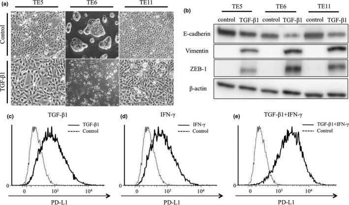
Figure 5.

ZEB1 and PD‐L1 expression and morphological changes in esophageal squamous cell carcinoma cell lines induced by TGF‐β1. TE5, TE6 and TE11 cells were treated by TGF‐β1 for 96 h. (a) Morphological changes of TE5, TE6, and TE11 treated by TGF‐β1 for 96 h. (b) Western blot analysis of the indicated proteins in cells treated by TGF‐β1 for 96 h. (c–e) FACS analysis of surface expression of PD‐L1 in TE6 cells co‐treated with TGF‐β1 and IFN‐γ as indicated.
