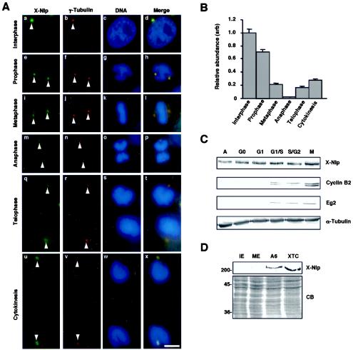
FIG. 2.
Localization of X-Nlp at the centrosome is cell cycle dependent. (A) Immunofluorescence microscopy of methanol-fixed Xenopus A6 cells at different stages of the cell cycle, as indicated, costained with purified R1679 anti-X-Nlp (green) and anti-γ-tubulin (red) antibodies. DNA was stained with Hoechst 33258 (blue), and merged images are shown. Arrowheads indicate positions of centrosomes/spindle poles. (B) The abundance of X-Nlp at a pair of interphase centrosomes (given an arbitrary value of 1.0) was compared with that at the two spindle poles (added together) in different stages of mitosis by quantitative fluorescence imaging as described in Materials and Methods. The centrosomes/spindle poles in 20 cells were analyzed for each bar of the histogram, and error bars represent standard deviations. (C) Xenopus A6 cells were synchronized in different stages of the cell cycle, as indicated (A, asynchronous), with the procedures described in Materials and Methods. Cell extracts were separated by SDS-PAGE and Western blotted for X-Nlp (purified R1679 antibodies), cyclin B2, Eg2, and α-tubulin. (D) Interphase (IE), and metaphase II (ME)-arrested Xenopus egg extracts as well as extracts of A6 and XTC cells were Western blotted for X-Nlp protein (purified 1679 antibodies, upper panel). Equal protein loading was confirmed by Coomassie blue (CB) staining of an equivalent SDS-polyacrylamide gel (lower panel). The positions of molecular size markers are indicated on the left (in kilodaltons).

