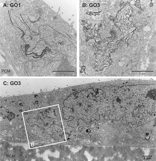
Fig. 4.

Internalization of GO by undifferentiated Caco-2 cells II. TEM micrographs of undifferentiated Caco-2 cells grown on permeable supports. Caco-2 after exposure to 20 µg/ml GO1 (A) or GO3 (B, C) for 24 h; C composed image of TEM micrographs showing parts of a Caco-2 cell with intracellular accumulation of GO3 sheets; B higher resolution image of framed area in C. NC nucleus, PCM polycarbonate membrane. Scale bars 2 µm
