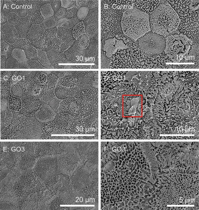
Fig. 7.

Cell surface morphology of differentiated Caco-2 cells. Scanning electron microscopy (SEM) images of differentiated Caco-2 cells grown on permeable membrane supports. Images show control cells without GO exposure (A, B) and cells after exposure to 20 μg GO1/ml (C, D) or 20 μg GO3/ml (E, F) for 24 h. Only a few GO1 sheets could be identified on the brush border surface (highlighted by red box), whereas no GO3 sheets could be clearly identified. For further SEM images see Additional File 1: Figure S6
