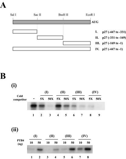
FIG. 2.
Determination of the PTB-binding site on the p27 IRES. (A) Schematic diagram of the p27 RNAs used in the various competition experiments. AUG denotes the initiator AUG codon. (B, i) Competition of RNAs for interaction with PTB. Competition experiments were carried out with purified PTB4 (100 ng), 32P-labeled RNA corresponding to probe IV of panel A (lane 1), and cold competitor RNAs (probe I [lanes 2 and 3], probe II [lanes 4 and 5], probe III [lanes 6 and 7], and probe IV [lanes 8 and 9]) in a 5-fold (lanes 2, 4, 6, and 8) or 50-fold (lanes 3, 5, 7, and 9) molar excess. (ii) Binding of PTB to various regions of the p27 5′NTR. UV cross-linking experiments were performed with 10 ng (lanes 1, 3, 5, and 7) or 50 ng (lanes 2, 4, 6, and 8) of purified PTB4 and 32P-labeled RNAs corresponding to probe I (lanes 1 and 2), probe II (lanes 3 and 4), probe III (lanes 5 and 6), and probe IV (lanes 7 and 8).

