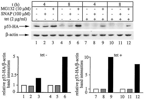
FIG. 6.
Proteasomal degradation of p53 is not impaired by NO. H1299 cells were plated for 24 h in tetracycline (tet)-free media to express p53-HA. Tetracycline (2 μg/ml) was then added back to half of the cells (lanes 7 to 12) to shut off the transcription of p53-HA cDNA. The cells were either left untreated (lanes 1, 4, 7, and 10), treated with 100 μM SNAP for 4 h (lanes 2 and 8) or 8 h (lanes 5 and 11), or treated with 10 μM MG132 for 4 h (lanes 3 and 9) and 8 h (lanes 6 and 12), respectively. When necessary, a fresh bolus of SNAP was added after 4 h. Cell lysates were subjected to Western blotting with HA (top) and β-actin antibodies (bottom). The immunoreactive bands were quantified by densitometric scanning. The p53-HA/β-actin ratios are plotted at the bottom. t, time.

