FIG. 2.
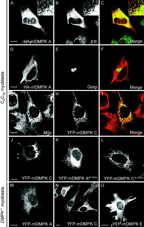
Mouse DMPK A and C localize to the ER or mitochondria. To assign subcellular DMPK location, confocal images were taken of fluorescent products in C2C12 myoblast cells 24 h after cotransfection with HA-tagged or YFP-tagged mDMPK isoforms A and C and various organellar markers. (A to C) HA-mDMPK A colocalized with a GFP-ER marker containing the calreticulin ER-targeting signal and the KDEL ER retention signal. The insert shows an enlargement of the peripheral ER network. (D to F) Fluorescent visualization of cotransfected HA-mDMPK A (D) and a GFP-Golgi (E) marker shows that mDMPK A is not targeted to the Golgi. (G to I) YFP-mDMPK C localized to mitochondria, as shown by staining with an anti-cytochrome c oxidase antibody. (J) Strong overexpression of YFP-mDMPK C leads to a cytosolic location in addition to perinuclear mitochondrial clustering. (K and L) Localization of kinase-dead mutants YFP-mDMPK A(K100A) and C(K100A) to the ER and mitochondria, respectively. (M and N) Adenovirus gene transfer of YFP-mDMPK A and C into DMPK−/− myoblasts result in ER and mitochondrial localization, respectively. (O) Expression of YFP-mDMPK E in DMPK−/− myoblasts gives a cytosolic distribution. Bars, 10 μm.

