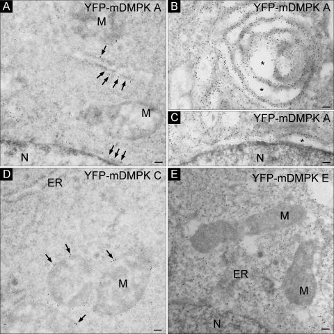
FIG. 3.
Mouse DMPK A and C localize to ER membranes or the MOM. Immuno-EM of YFP-mDMPKs A, C, or E in transiently transfected N2A cells visualized with an anti-GFP antibody followed by incubation with protein A complexed to 10-nm gold particles is shown (A). YFP-mDMPK A localized to ER and NE structures. (B and C) Overexpression caused abnormal ER membrane stacks (B) next to dilated ER and NE structures (B and C, asterisks). (D and E) YFP-mDMPK C localized exclusively to the MOM (D), whereas a cytosolic localization was observed for YFP-mDMPK E (E). M, mitochondrion; N, nucleus. Bars, 100 nm.

