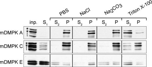
FIG. 4.
Mouse DMPK A and C are tail-anchored proteins. Western blots of pellet (P) and supernatant (S1 and S2) fractions of N2A cells transfected with mDMPK A, C, or E after phosphate-buffered saline (PBS), high-salt, alkaline Na2CO3, or Triton X-100 extraction of 100,000 × g pellets. N2A cells were grown, transfected, and fractionated into supernatant and pellet fractions before extraction as described in Materials and Methods. DMPK was visualized with an anti-DMPK antibody. inp., input; S1/S2 and P, supernatant and pellet fractions after 100,000 × g centrifugation. Asterisks mark doublet DMPK signals, and brackets indicate C-terminally truncated mDMPK products as a result of nonenzymatic breakdown (see the text).

