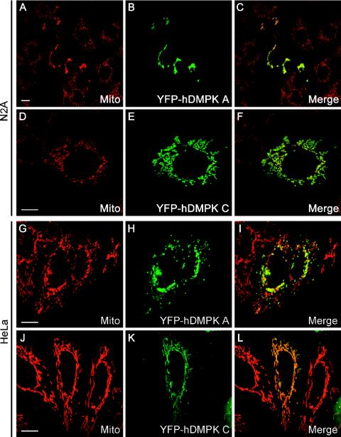
FIG. 7.
Human DMPK A and C localize to mitochondria and alter mitochondrial distribution. Confocal fluorescence microscopy analysis of YFP-hDMPK A and C in transiently transfected N2A (A to F) and HeLa (G to L) cells. YFP-hDMPK A localized at mitochondria, stained by an anti-cytochrome c oxidase marker, which accumulated in the nuclear periphery. YFP-hDMPK C also localized to mitochondria (D to F and J to L) but did not show the accumulation near the nucleus as observed for hDMPK A (B and H). Note that mitochondria devoid of hDMPK A remain unclustered (H). Bars, 10 μm.

