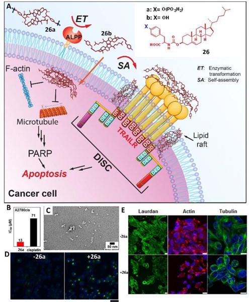
Fig. 12.

(A) EISA of the tyrosine-cholesterol conjugate activates extrinsic and intrinsic cell death signaling. (B) IC50 of 26a and cisplatin against A2780cis cell lines for 48 h. (C) TEM images of 24a (100 μM) treated with ALP (1 U/mL) in PBS (pH7.4) after 24 h; bar is 50 nm. (D) Confocal images of A2780cis cells treated with anti-DR5 without or with the addition of 26a (12.5 μM) for 24 h. Scale bar = 30 μm. (E) Confocal images of A2780cis cells stained with laurdan (10 μM), Alexa Fluor 633 phalloidin (F-actin, red), and Hoechst (nuclei, blue) or tubulin tracker (green) without or with the addition of 26a (12.5 μM) for 12 h (scale bar is 10 μm). Adapted with permission from ref. 36. Copyright 2016, American Chemical Society.
