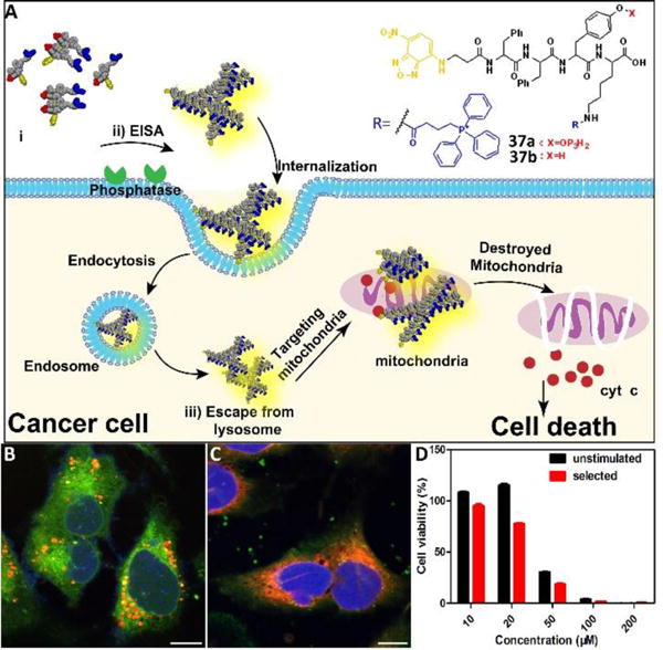
Fig. 16.

(A) Illustration of enzyme-instructed self-assembly for targeting mitochondria and inducing the death of cancer cells. (B) CLSM images of Saos-2 cells treated with D-37a (50 μM) for 4 h and then stained with Lyso-Tracker. Scale bar is 10 μm. (C) CLSM images of Saos-2 cells treated with D-37a (50 μM) for 4 h and then stained with Mito-tracker. Scale bar is 10 μm. (D) Cell viability of unstimulated Saos-2 cell line or selected Saos-2 cell line (after five weeks treatment of the precursors with gradually increase concentrations) incubated with D-37a at different concentrations for 48 h. Adapted with permission from ref.43. Copyright 2016, American Chemical Society.
