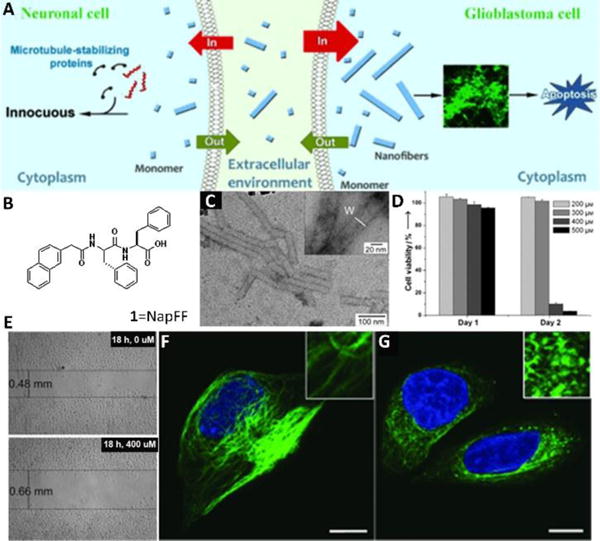
Fig. 2.

(A) Proposed mechanism for the selective inhibition of the growth of glioblastoma cells by the self-assembled nanofibers of 1. (B) Molecular structure of 1. (C) Negatively stained TEM images of a solution of 1 (400 μM) in PBS buffer. (D) Cell-viability assays of HeLa cells after treatment for 24 and 48 h with 1 at different initial concentrations. (E) Cell-migration assay of HeLa cells treated with 1. Confocal images of tubulin-stained T98G cells treated with a medium containing 1 at a concentration of 0 (F) or 400 μM (G) for 24 h. Insets are images that have been enlarged further by a factor of 3. Scale bar: 10 μm. Adapted with permission from ref. 19. Copyright 2013, Wiley-VCH.
