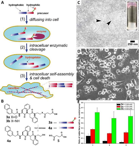
Fig. 4.

(A) Schematic intracellular formation of nanofibers that leads to hydrogelation and cell death. (B) The chemical structures and graphic representations of the precursor (3a), the control molecule (3b), and the hydrogelator (4a). (C) TEM of the hydrogels formed by the dead HeLa cells after culturing with 3a for three days (arrows indicated the nanofibers formed by 4a). (D) Optical images (x100) of the growth of HeLa cells after being cultured in the media containing 0.08 wt% of 3a in 1 day. (E) MTT assays of HeLa cells treated with 3a at concentrations of 0.08 wt%, 0.04 wt%, and 0.02 wt%. Adapted with permission from ref. 23. Copyright 2007, Wiley-VCH.
