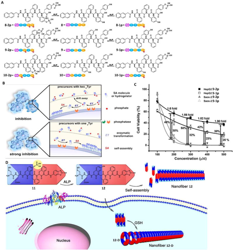
Fig. 6.

(A) Molecular structures of the precursors (8-2p, 9-2p, 10-2p, 8-1p, 9-1p, and 10-1p) that have one or two phosphotyrosine residues and the corresponding self-assembling D-peptides (i.e., hydrogelators 8, 9, and 10). (B) EISA to inhibit cancer cells. (C) 48-hour cytotoxicity of 9-2p and 9-1p on HepG2 and Saos-2 cells at different concentrations. The viability differences between the two cell lines are labelled as well. (D) Schematic illustration of ALP-directed self-assembly of 11 into nanofiber 12 in extracellular environment, and GSH-controlled condensation of 12 to yield the cyclic amphiphilic dimer 12-D, which self-assembles into nanofiber 12-D in an intracellular environment. Blue parts indicate the hydrophilic structures, and red parts indicate the hydrophobic structures. Adapted with permission from ref. 45, 46 and 47. Copyright 2016, American Chemical Society (ref. 26 and 28). Copyright 2016, Wiley-VCH (ref. 27).
