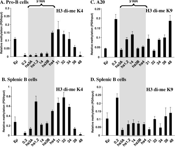
FIG. 4.
Reciprocal association of di-me K4 H3 and di-me K9 H3 at sequences flanking the extended 3′ RR and real-time PCR analysis of di-me K4 H3 and di-me K9 H3 modifications. Shown is the enrichment of di-me K4 H3 in pro-B cells (A) and in splenic B cells (B) and the enrichment of di-me K9 H3 in A20 cells (C) and in splenocytes (D).

